Most drugs work by fitting into a pocket on the surface of a protein. In this walkthrough we look at Abl kinase, the enzyme switched on in chronic myeloid leukaemia, in a structure (PDB 2HYY) solved together with the drug imatinib. First we find the pocket and the residues that hold the drug, then we take the drug out and dock it back, to see whether docking finds the right place. Every side chain and ligand we look at is drawn as sticks, coloured by element: carbon grey, nitrogen blue, oxygen red, sulfur yellow.
1. Load the structure and show its fold
Open ProKyria with the button above, or type 2HYY into Load a Structure in the Proteins tab and press Load. The file holds four copies of the kinase domain (chains A to D), each with an imatinib molecule bound.
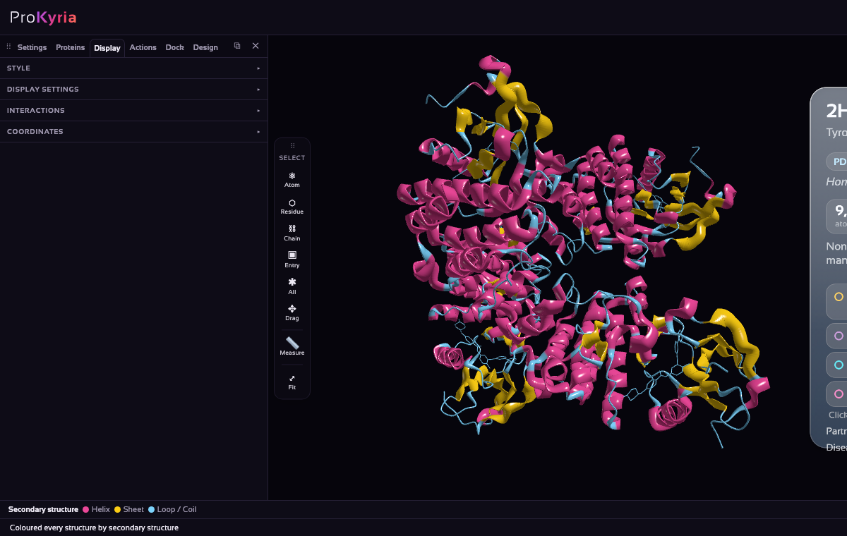
In Display › Style, choose Ribbon and colour by Sec. structure. With nothing selected, this applies to the whole structure: helices turn pink, strands yellow and loops light blue. Each kinase domain shows the classic two lobes, a small one of strands and a large one of helices, with the drug-binding cleft between them.
2. Show the drug and the residues round it
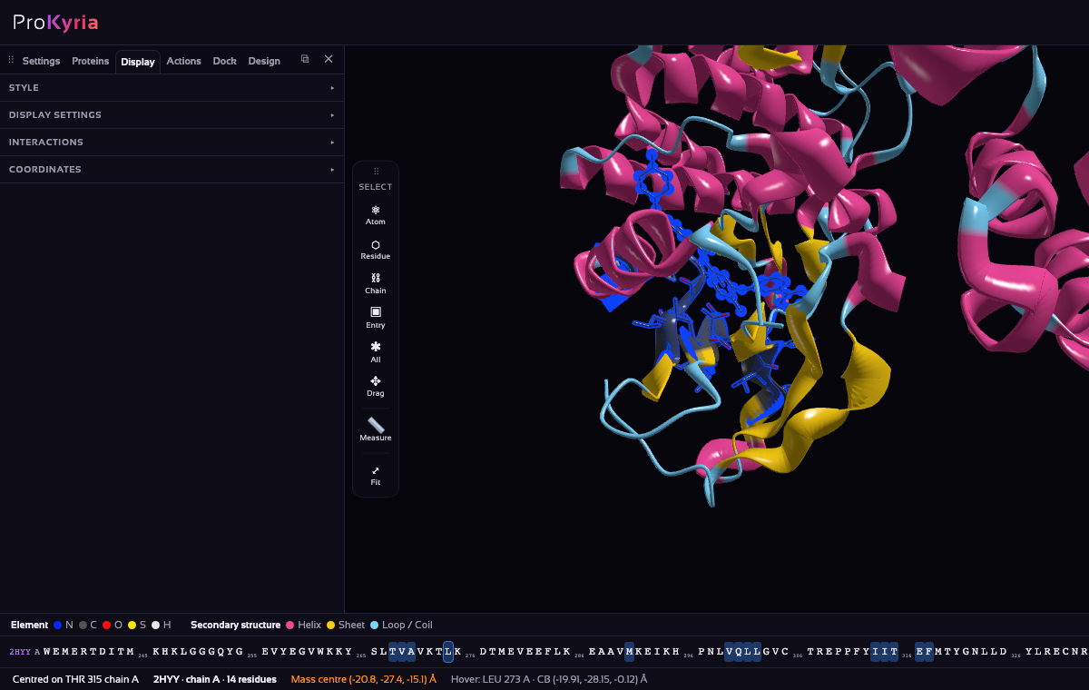
Choose Residue on the tool wand's Select buttons and click imatinib in chain A (residue STI 600, sitting in the cleft). In Display › Style, choose Stick and colour it by Element.
Now take the residues that touch it. Right-click the drug and, on the Select within row, leave the distance at 5 Å and press Residues. Everything within 5 Å is added to the selection. Choose Stick and colour by Element again, so their side chains show against the ribbon. Most of them are carbon-rich and hydrophobic, which is why a flat, greasy drug binds here.
3. Find the drug's hydrogen bonds
A drug is held by its contacts with the protein, and hydrogen bonds are the most specific of them. Select imatinib again (Residue on the tool wand, then click it). In Display › Interactions, tick Hydrogen bonds, π-stacking and Only around the selection, with within a chain on (the drug belongs to chain A). Dashed lines appear between the drug and the residues it bonds to: blue for hydrogen bonds, green for π-stacking. The summary under the boxes counts them.
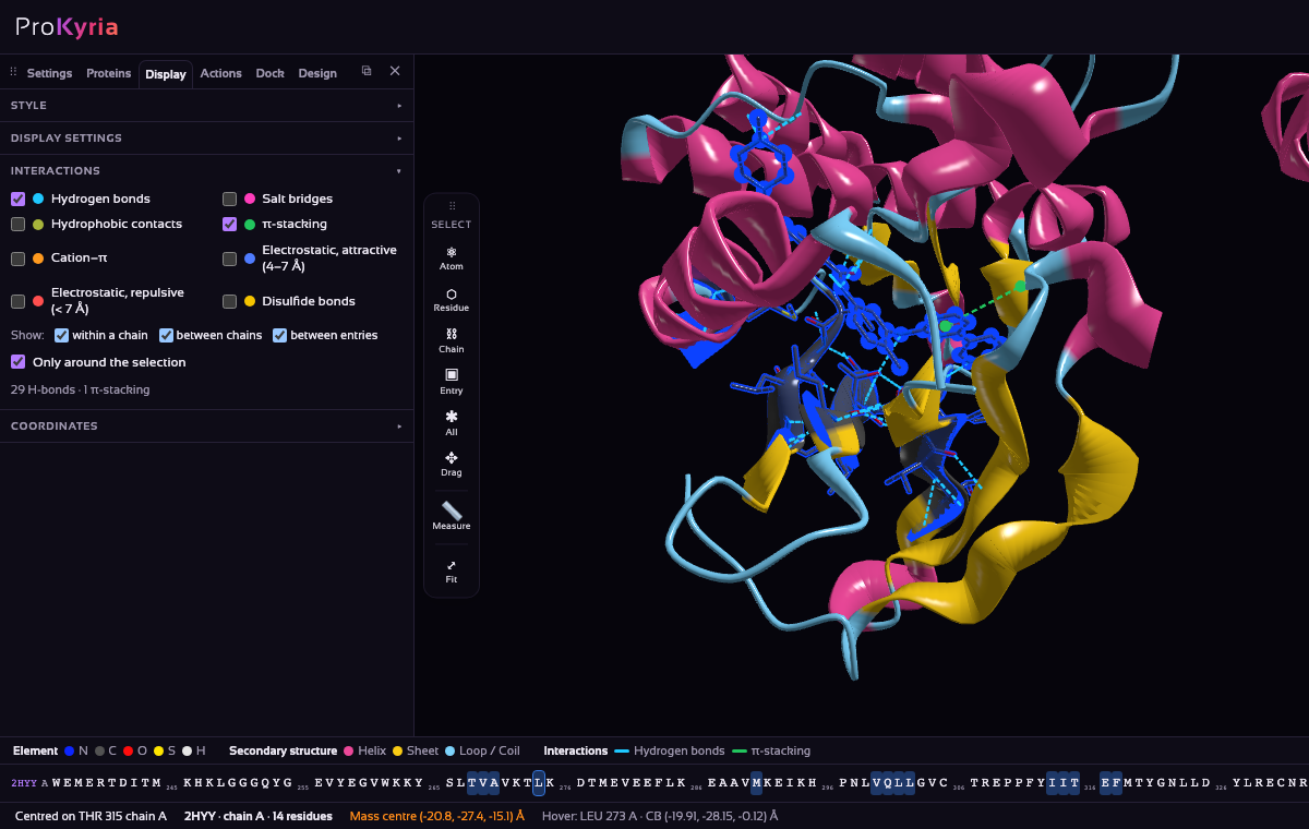
Turn on the sequence bar (Display › Display settings › Amino acid sequence) and hover over the residues at the ends of the lines to read their names. Imatinib makes eight hydrogen bonds:
- Met318, the hinge: its backbone N–H bonds to the drug's pyridine nitrogen. Nearly every kinase inhibitor makes a hinge bond like this one.
- Thr315, the gatekeeper: its side-chain O–H bonds to the drug's amine N–H. In the resistance mutation T315I, isoleucine has no O–H, so this bond is lost, and its bulkier side chain gets in the drug's way.
- Glu286 (on the αC helix) and the backbone of Asp381 (the DFG motif) hold the amide in the middle of the drug.
- Ile360 and His361: their backbone C=O groups bond to the charged N–H of the piperazine ring at the drug's far end.
The green line is a π-stack between one of the drug's rings and Tyr253 in the glycine-rich loop. Tick Hydrophobic contacts as well to see how the rest of the pocket (Phe382, Ile313, Phe317 and others) wraps the drug's flat rings.
4. Detect the pockets
Run Geometric Cavity in Actions: open the Actions tab and, under Pocket Detection, leave the method on Geometric Cavity and press Run. After a few seconds each pocket appears as a coloured cloud, and the list gives its size, a score, and the residues that line it.
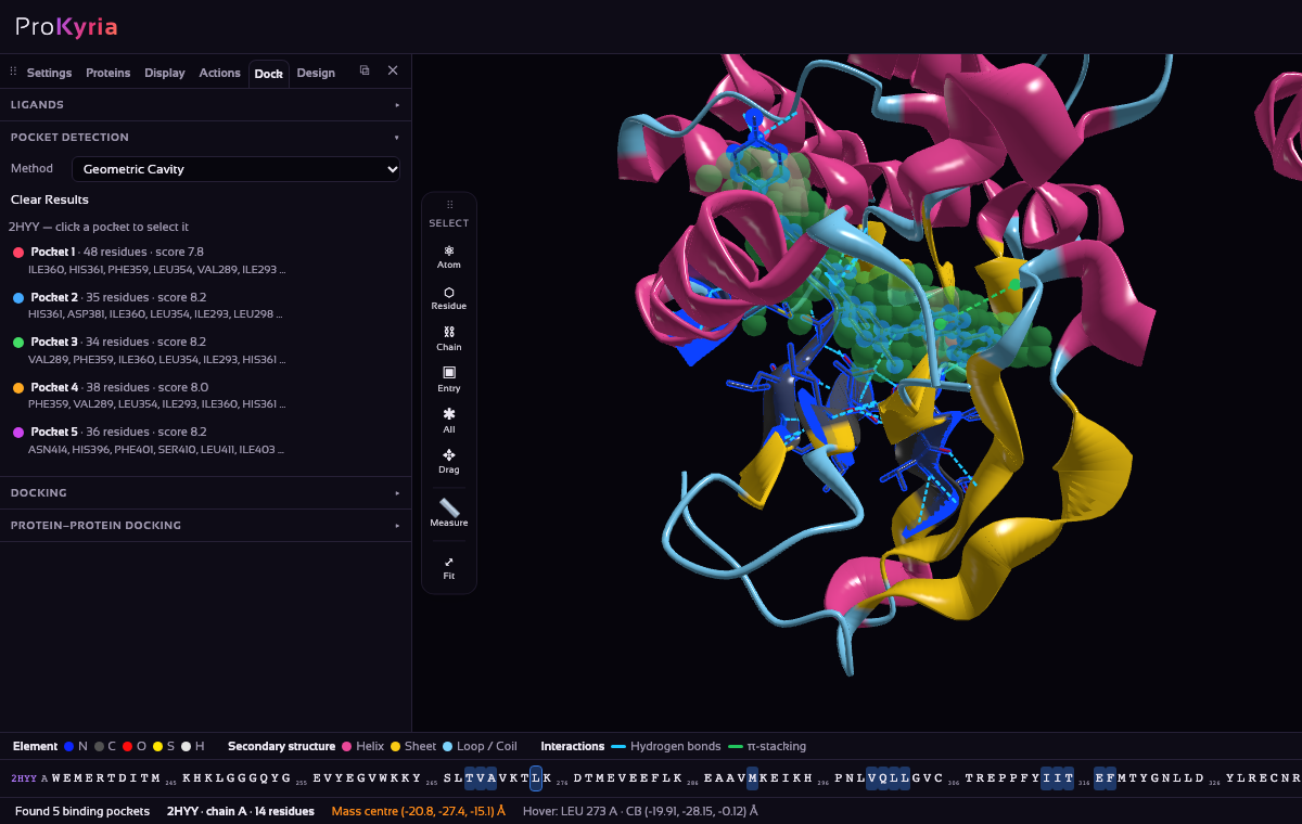
Find the pocket lined by Thr315, Glu286 or Asp381 and click it to select its residues: it is the cleft where imatinib sits, found without being told where to look.
Want a second opinion? Switch the method to Alpha-Sphere and run it again. It describes pockets as clusters of empty spheres and adds a druggability estimate. See how alpha-sphere detection works.
5. See which drugs are already known
Still in the Actions tab, under Ligands, pick the protein in Known ligands of. ProKyria looks up its UniProt entry (ABL1, P00519) and asks several sources at once: Open Targets for approved and clinical-stage drugs, the PDB for molecules bound in other structures of the protein, and UniProt for annotated binding sites.
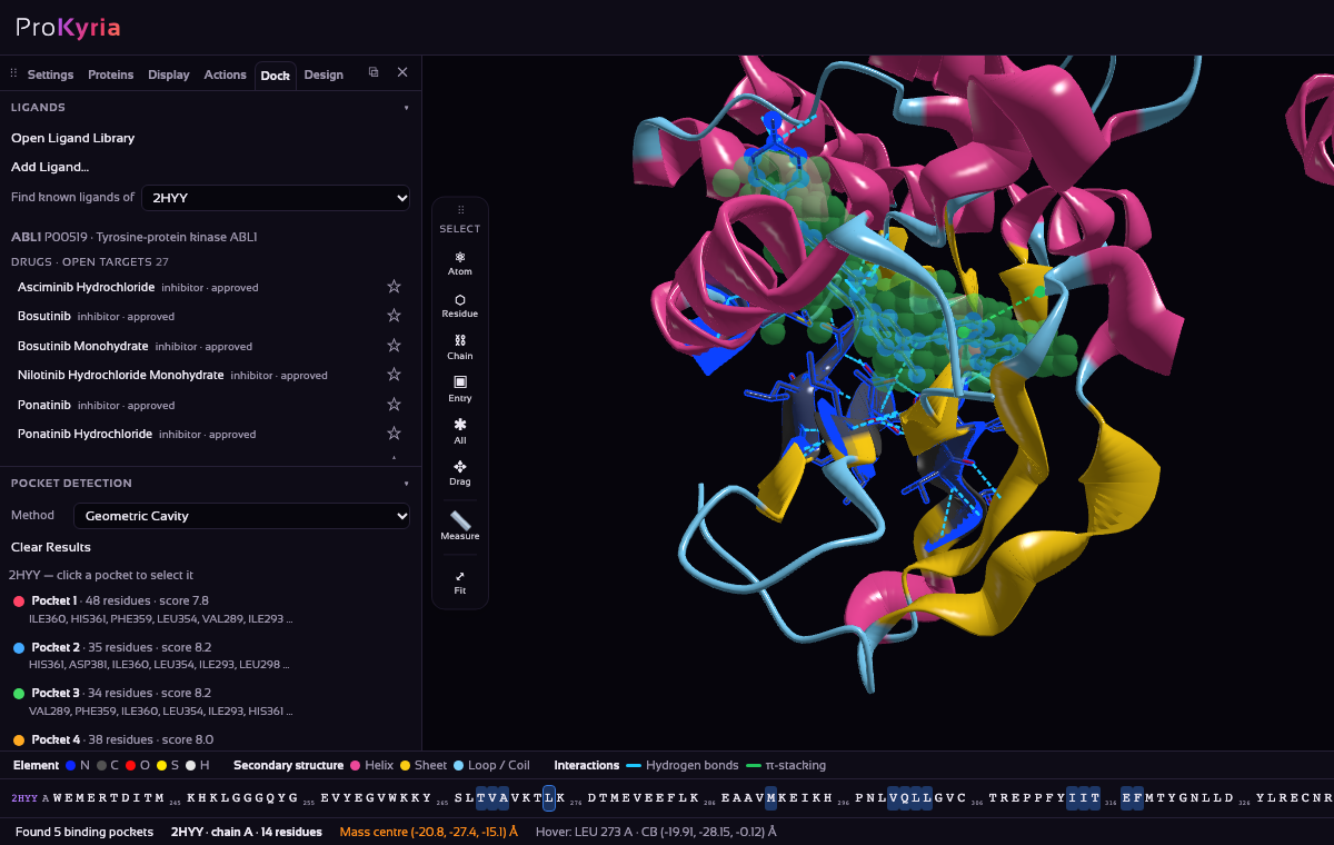
For Abl the list includes the approved kinase inhibitors asciminib, bosutinib, nilotinib and ponatinib. Click a row to load the molecule, or its star to keep it in your ligand library.
6. Take the drug out
Now check that docking can find this binding mode by itself. This is redocking: you take the answer out and see whether the method puts it back. To keep the calculation quick, work with one copy of the kinase. In the Proteins tab, expand the entry in Loaded Entries with ▸ to see its chains, then right-click chains B, C and D and choose Delete chain.
Then remove chain A's imatinib, otherwise it would fill the pocket: choose Residue on the tool wand, click the drug, and press Delete (or right-click it and choose Remove residue). Everything can be undone with Ctrl/⌘+Z.
7. Load the ligand
In Load a Structure, set Source to SMILES and type imatinib, or paste its SMILES string. ProKyria fetches a 3D model of the molecule from PubChem, with hydrogens, and loads it as its own entry, drawn as sticks by element. You can also upload an .sdf or .mol file.
8. Choose the site
Run Geometric Cavity in Actions again, now on the single chain. The first pocket becomes the docking Site, and a box shows the region that will be searched. Check it holds Thr315 and Met318, still drawn as sticks.
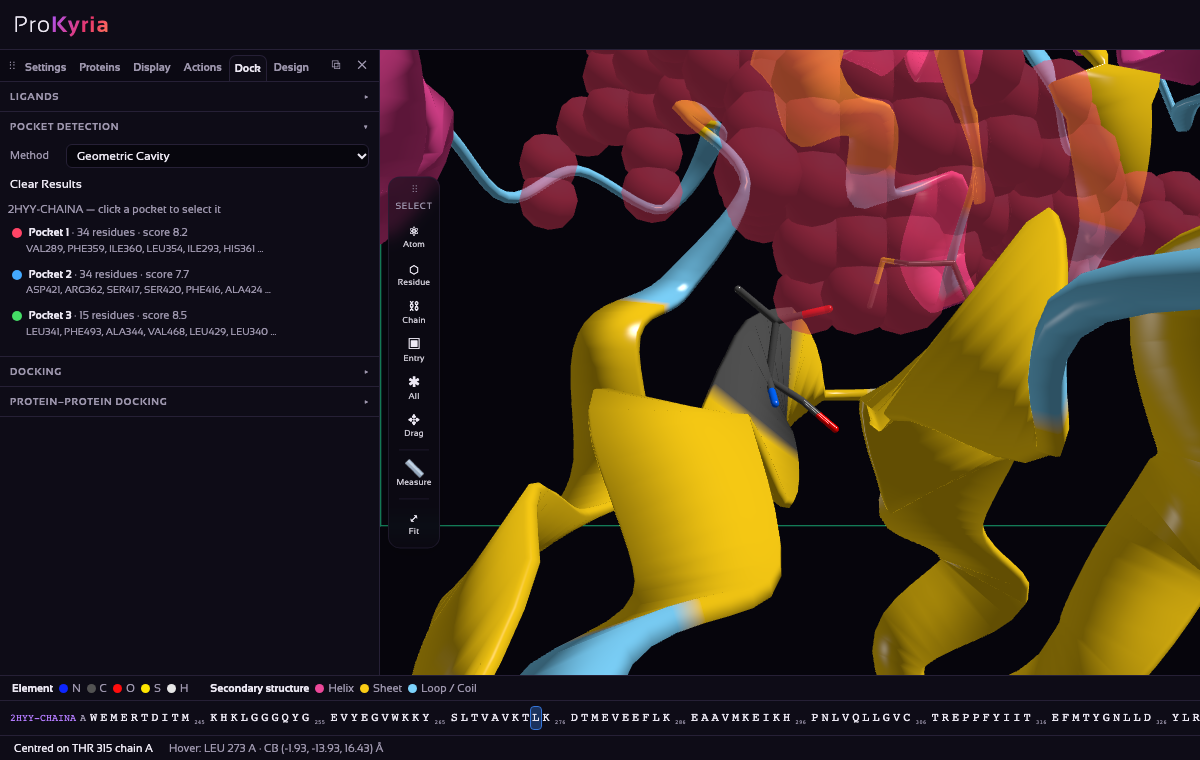
In the Docking section, check that the Receptor is the kinase and the Ligand is imatinib, and keep the method on Built-in · Vina-style scoring. A few settings are worth knowing:
- Site: a detected pocket, the residues you've selected, or the whole protein ("blind" docking, which is slower).
- Poses: how many different binding modes to return.
- Exhaustiveness: how hard to search. Higher values are slower but less likely to miss the best pose.
- Box padding: how far the search box extends beyond the site.
9. Run the docking
Press Run Docking. It runs entirely in your browser, so nothing is sent to a server. A small molecule in a pocket usually takes seconds to a minute.
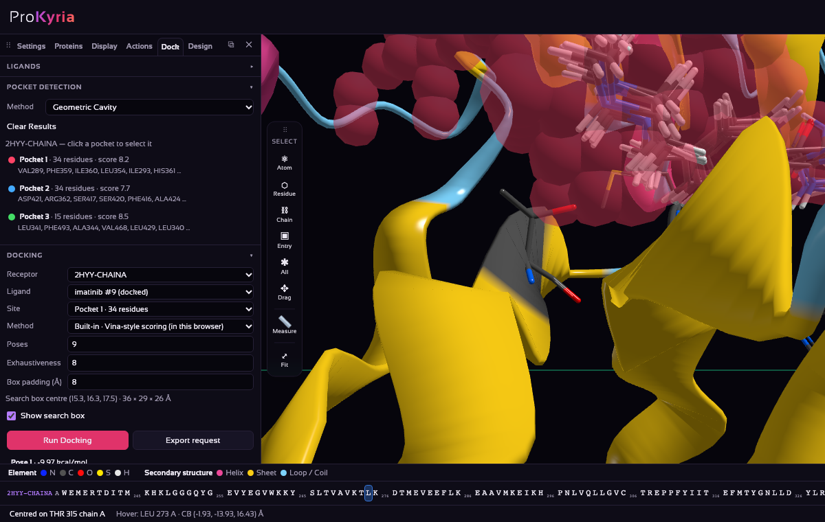
Each pose is added as a new entry ("imatinib #1", "#2", …) with a predicted binding score in kcal/mol, where more negative means a better fit. Hide every pose but the best with its eye button in Loaded Entries. The best pose is drawn as sticks by element, like everything else in the pocket.
10. See how the pose is held
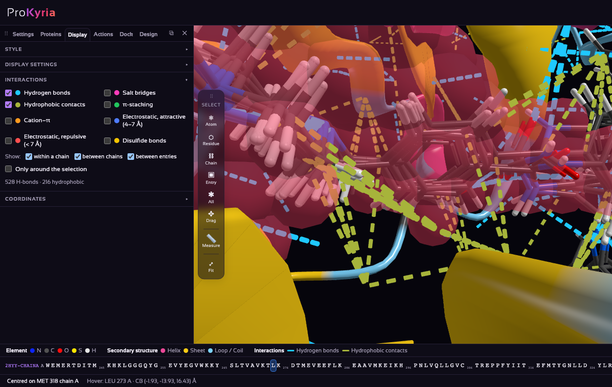
In Display › Interactions, untick Only around the selection, keep Hydrogen bonds on, tick Hydrophobic contacts, and keep between entries on: the docked pose is its own entry. Dashed lines join the pose to the protein: blue for hydrogen bonds, olive for hydrophobic contacts.
Compare them with the crystal's in step 3. A good pose makes the same key bonds: to the backbone of the hinge Met318 and the side chain of the gatekeeper Thr315, and ideally to Glu286 and Asp381. A pose with a good score but none of these bonds is probably wrong.
11. Check the answer
Load 2HYY again and use Actions › Superposition to overlay the original crystal structure on your receptor. Its imatinib is drawn as sticks by element too, so colour the copy's ribbon Solid grey to tell the two apart. If your pose sits where the crystal's imatinib does, the set-up is sound, and you can dock molecules whose binding mode you don't know.
What next
- Try a known drug from step 5 instead of imatinib, and compare its pose and score.
- Read how geometric pocket detection and small-molecule docking work, and what they can miss.
Docking scores are fast estimates, not measured affinities. They're most useful for ranking poses of one molecule, or for comparing similar molecules in the same pocket. Always look at the pose itself: does it make sensible hydrogen bonds, and does it fill the pocket? Pocket detection looks for buried cavities, so it works best on deep pockets like this kinase cleft.