Whether you're designing a protein or interpreting a patient's variant, the first question about a mutation is often simple: does the new side chain fit? ProKyria lets you mutate any residue in place, tells you what kind of change it is, and shows where the new side chain would bump into its neighbours.
We'll use crambin (PDB 1CRN), a small plant protein of 46 residues, held together by three disulfide bonds.
1. Load and colour by element
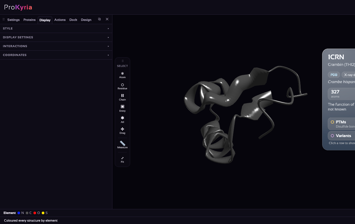
Load 1CRN. In Display › Style, choose Ribbon and colour by Element. With nothing selected, the style applies to the whole protein: the ribbon follows the backbone, and the atoms you draw later get the standard colours, carbon grey, nitrogen blue, oxygen red and sulfur yellow.
2. Select a residue and show it as sticks
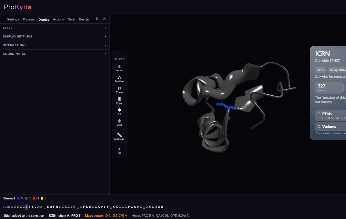
Choose Residue on the tool wand's Select buttons, then click a residue on the ribbon: here proline 5. The bottom bar names what you hover over, and the selection shows in blue. With the residue selected, click Stick in Display › Style. Only that residue is drawn as sticks, by element, so you can see its side chain and its neighbours before you change it.
3. Pick it for mutation
Right-click the residue and choose Mutate residue.
4. Choose the new amino acid
The Mutate Residue window shows the residue you picked. Choose the new amino acid under To, and ProKyria immediately lists what kind of change it is.
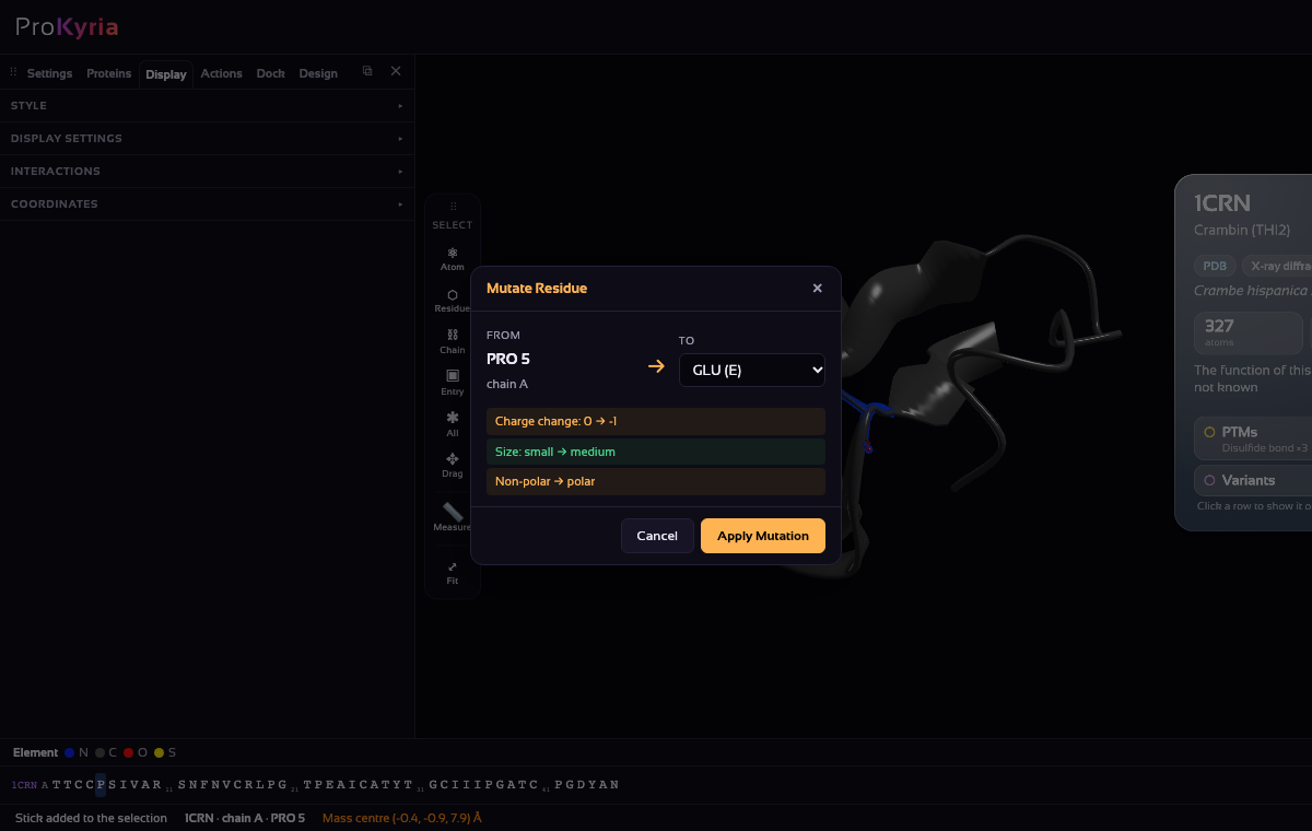
For proline to glutamate it notes three things: a charge change (0 → −1), a size change (small → medium) and non-polar → polar. Other mutations bring other notes. Introducing a proline may break a helix or strand, losing a glycine reduces backbone flexibility, and changing a cysteine may break a disulfide bond. Changes are coloured by how serious they're likely to be.
5. Apply it and look for clashes
Press Apply Mutation. ProKyria builds the new side chain onto the backbone and checks it against every nearby atom.
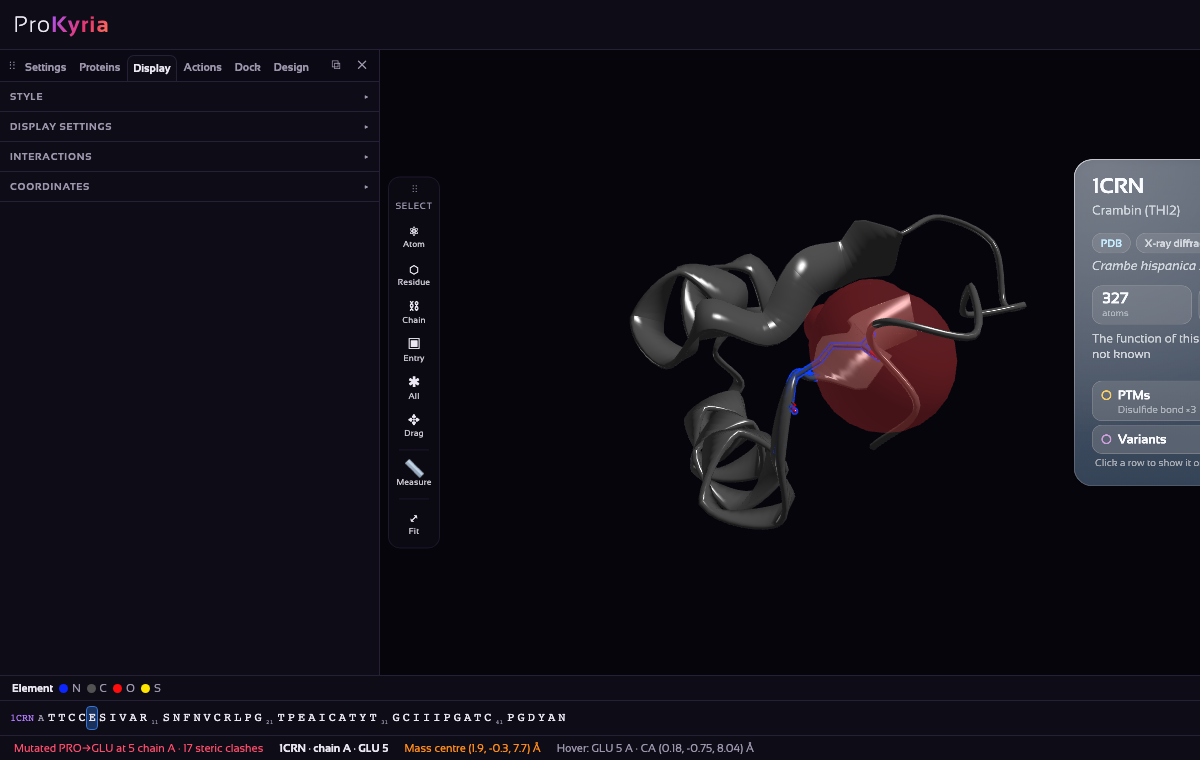
Red spheres mark steric clashes, atoms closer together than they can physically be, and the bottom bar counts them (17 here). Many clashes in a packed region suggest the new side chain doesn't fit without the structure rearranging. That's a hint the mutation could be destabilising.
The side panel now has a Mutations list. Undo Last and Revert All take changes back, and so does the usual Ctrl/⌘+Z.
What this does and doesn't tell you
This is a quick structural check, not a stability prediction. The new side chain is placed in a standard shape, and the protein doesn't relax around it. Some clashes would disappear if neighbouring side chains moved slightly. Use it to spot mutations that clearly can't fit, and to see which neighbours are affected. Use dedicated stability predictors or experiments for a real ΔΔG.
Next: run molecular dynamics on the mutant to see how its flexibility compares, or export it with Settings › Export › PDB.