A crystal structure is a single snapshot, but proteins are always moving. Loops flex, domains hinge, termini wave. ProKyria can simulate these large-scale motions in a few seconds, in your browser, with a coarse-grained elastic network model. We'll colour crambin by how mobile its atoms were in the crystal, mark what holds it rigid, and see whether the simulation agrees.
1. Colour by flexibility
Load crambin, 1CRN, a small plant protein of 46 residues. In Display › Style, choose Ribbon and colour by B-factor. The B-factor records how much each atom was smeared out in the crystal, a measure of flexibility: blue is rigid, and warmer colours up to red are more mobile. The legend at the bottom shows the range.
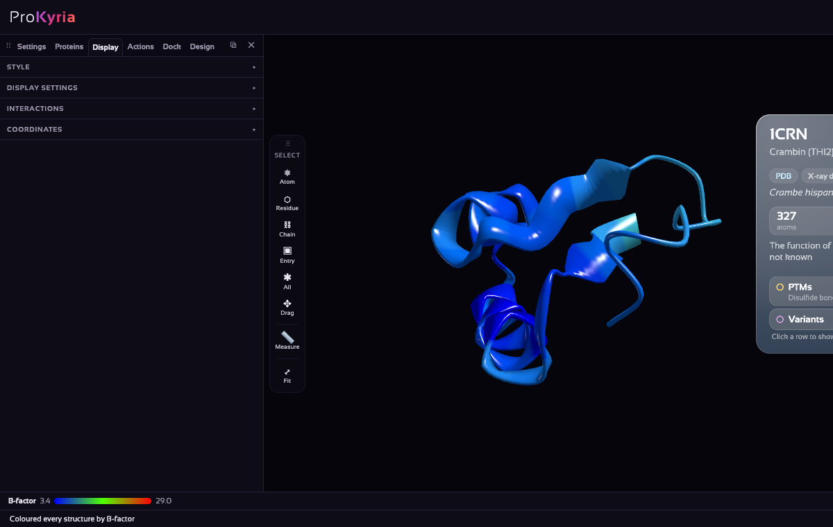
The warmest parts are the N-terminal Thr1, Tyr29 and the loop from Gly37 to Pro41.
2. Mark what holds it together
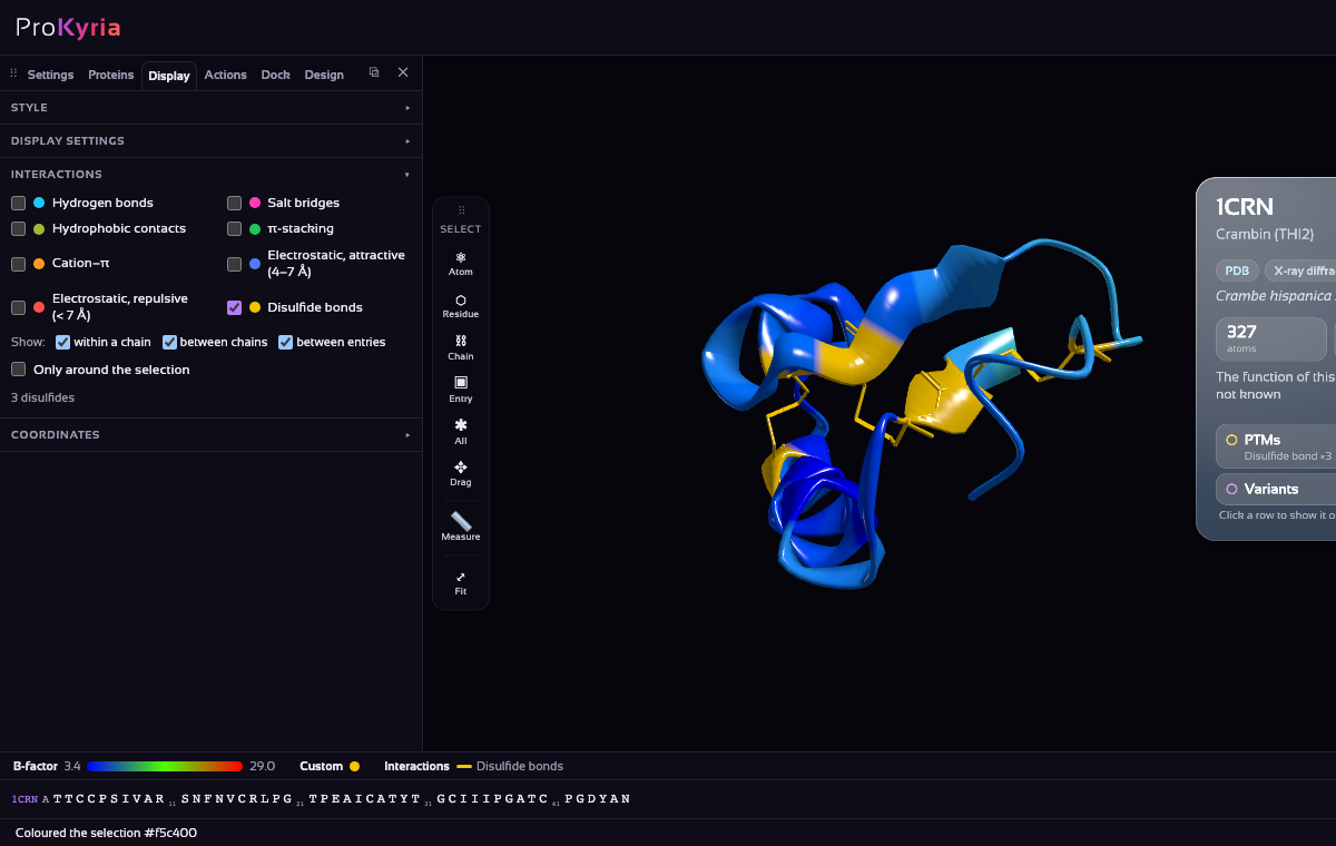
Crambin is stapled by three disulfide bonds: Cys3–Cys40, Cys4–Cys32 and Cys16–Cys26. Turn on the sequence bar (Display › Display settings › Amino acid sequence), click C3, and Ctrl/⌘-click the other five cysteines (C4, C16, C26, C32 and C40). Show them as Stick, with the Solid colour chip set to yellow.
Then, in Display › Interactions, tick Disulfide bonds: a dashed line joins each pair. Cys4, Cys16 and Cys26 are among the most rigid residues in the protein; only Cys40, beside the mobile 37–41 loop, is warmer.
3. Run the simulation
Open Actions. Under Molecular Dynamics, the System line says what will be simulated: everything loaded, or just the selection if you've selected something, so clear the selection first (double-click empty space). Set the Temperature (300 K is room temperature) and press Run.
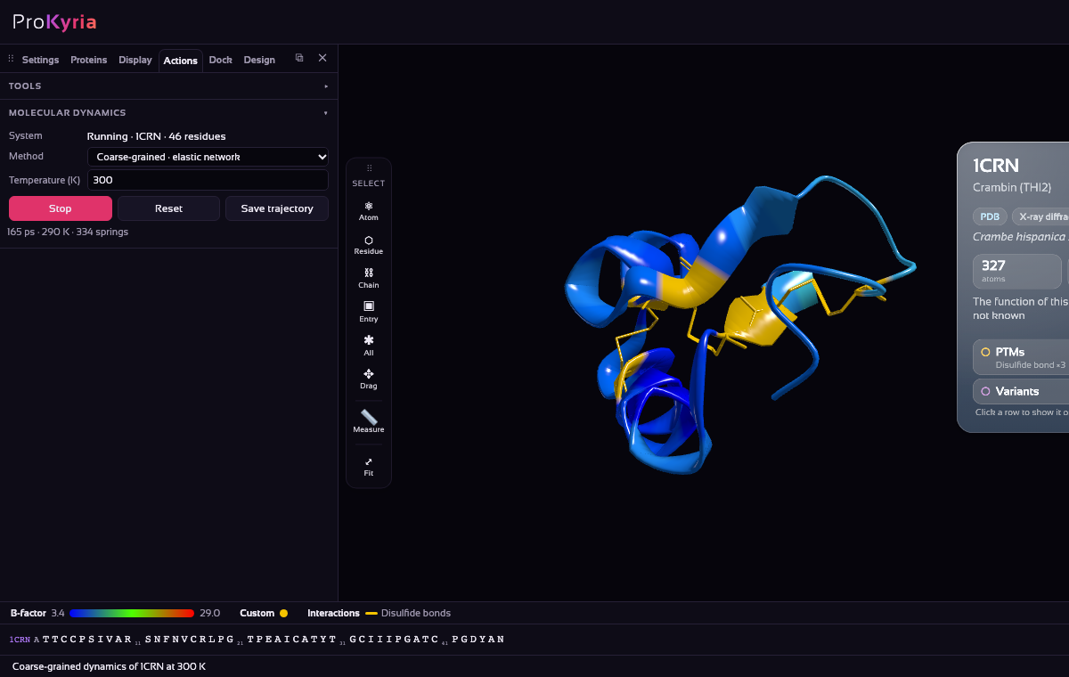
The protein starts moving straight away. The status line shows the simulated time in picoseconds, the current temperature and the size of the network. Change the temperature while it runs to see the motion grow or calm down. Press Stop when you've seen enough. The whole run is a single undo step, and a step in the Analysis log.
4. What you're seeing
Each residue is represented by one bead, joined by springs to every residue near it in the folded structure. The beads are then moved by random thermal kicks and friction. Tightly packed regions with many springs barely move, while loops and termini with few springs move freely. Watch the yellow cysteines stay put while the N-terminus and the 37–41 loop swing, as the B-factors said they would. That is why this simple model reproduces a protein's pattern of flexibility surprisingly well.
5. Keep the trajectory
Press Save trajectory to download the run as a multi-model PDB file, one model per frame. Open it in other programs, or load it again here and colour it by Variability, which shows how far each residue moved across the frames. Reset returns the structure to its starting coordinates.
Limits
An elastic network keeps the protein close to its starting fold. It shows natural fluctuations and collective motions, but not folding, unfolding or ligand binding, and it has no chemistry or solvent. Use it to explore flexibility and to compare variants. For atom-level dynamics you need an all-atom simulation package.
More detail: coarse-grained molecular dynamics in ProKyria.