A structure on its own doesn't tell you what a protein does, or which of its residues matter in disease. ProKyria can show that information next to the molecule. Every loaded protein can have an info card with data from its UniProt entry, and the entries on the card can be marked directly on the 3D structure. Here we use haemoglobin (PDB 4HHB) and end at the residue behind sickle-cell disease.
1. Load it and colour the chains
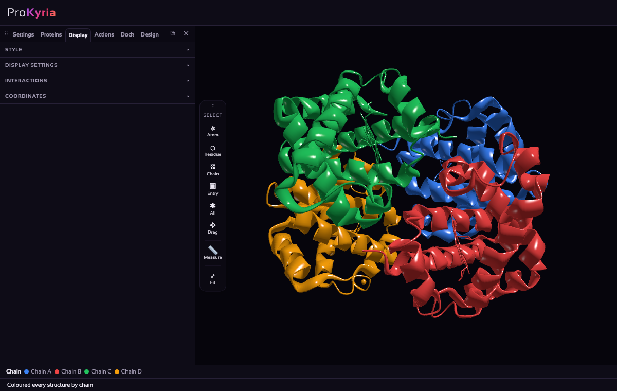
Load 4HHB, haemoglobin, the protein that carries oxygen in red blood cells. In Display › Style, choose Ribbon and colour by Chain. Chains A and C are the two α chains, B and D the two β chains; the legend at the bottom names each colour.
2. Highlight the haems
Each chain holds a haem, the iron-containing group that binds oxygen. Choose Residue on the tool wand's Select buttons, click one haem, and Shift-click the other three. Show them as Spacefill, coloured by Element: the iron at each centre is orange. Each haem sits in its own pocket, held by a histidine below the iron (His87 in α, His92 in β).
3. Turn on the info cards
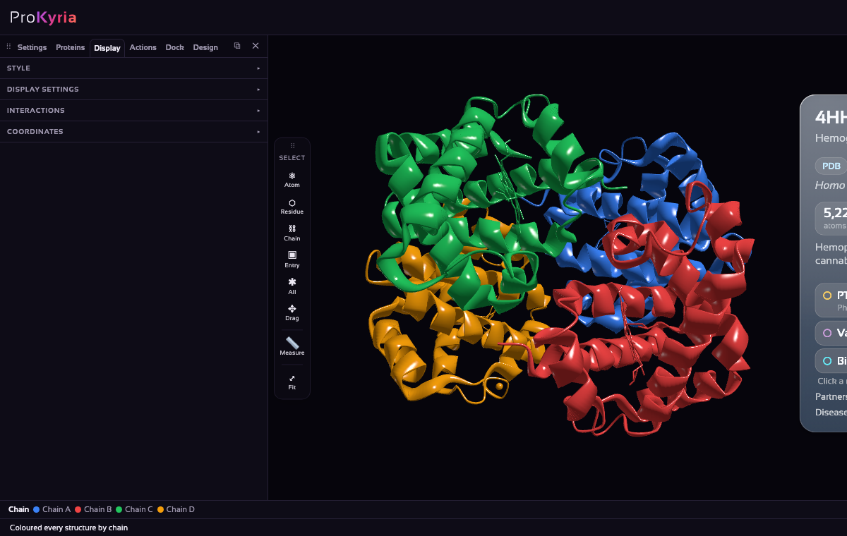
In the Display tab, tick Protein info cards. The card floats beside the protein and always faces you. It shows how the structure was determined (here X-ray diffraction at 1.74 Å) and the protein's size. It also shows what UniProt records: its function, post-translational modifications, variants, binding sites and associated diseases, such as forms of thalassaemia.
4. Put the annotations on the structure
Click the card's Variants row. Every residue with a known variant is selected, in the usual blue highlight, and drawn as sticks, its charged side chains coloured red (positive) or blue (negative). Click the row again to put them back. This shows at a glance whether disease variants cluster near the haem pockets, at the contacts between chains, or on the surface.
Drag the card to move it, and use its × to close it. Display › Protein info cards brings closed cards back.
5. Find the sickle-cell residue
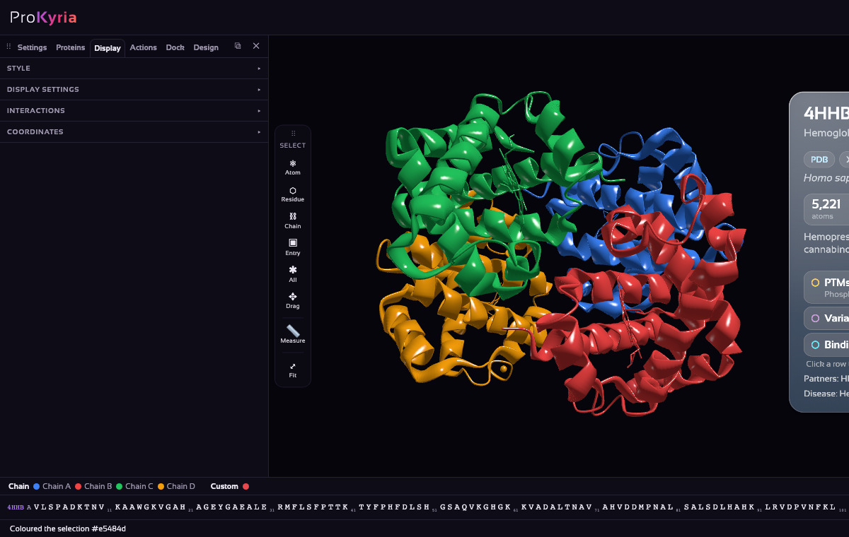
Sickle-cell disease comes from a single change in the β chain: glutamate 6 to valine. Turn on the sequence bar (Display › Display settings › Amino acid sequence) and, in chain B's row, click E6. Show it as Spacefill with the Solid colour chip set to red.
It sits on the outside of the β chain, in plain view. That is the point: a charged glutamate on the surface is harmless, but a valine there is a sticky, hydrophobic patch. In deoxygenated haemoglobin it fits a pocket on a neighbouring molecule, and the molecules line up into the long fibres that bend red cells into sickles. (UniProt counts from the initiator methionine, so its variant list calls this position Glu7.)
Tips
- To make the sickle-cell variant, right-click E6 and use Mutate residue to change it to VAL.
- Colour by Hydrophobicity to see the patch a valine would add to the surface.
- The card uses the UniProt entry linked from the PDB file. A structure without that link won't have one.
The annotations come from UniProt. Please credit it if you use them in your work.