Many proteins are assemblies of several chains. An antibody is the textbook example: two identical heavy chains and two identical light chains make the Y shape, with sugars attached to its stem. Here we take an intact antibody (PDB 1IGT) apart by colour: first every chain, then heavy against light, then the sugars.
1. Colour by chain
Load 1IGT. In Display › Style, choose Ribbon and colour by Chain. With nothing selected, the style applies to the whole structure.
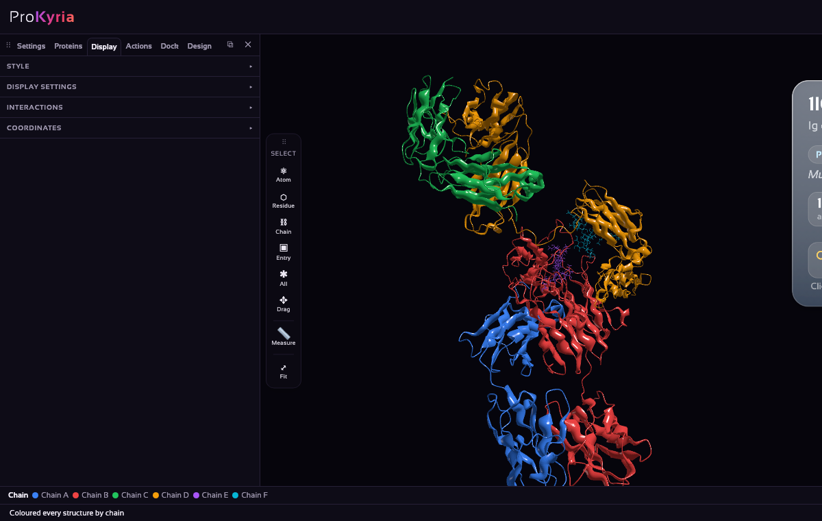
The legend strip at the bottom names each colour. Chains B and D are the heavy chains (444 residues each) and A and C the light chains (214 each). Follow one heavy chain from the tip of an arm down into the stem, then find the light chain alongside it in the arm.
2. Heavy against light
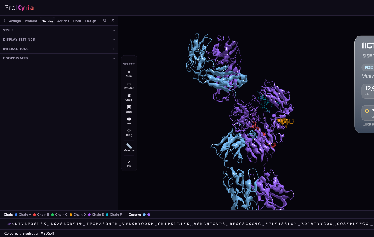
Colour the two kinds of chain apart. Choose Chain on the tool wand's Select buttons, click a light chain, and Shift-click the other. Choose the Solid colour chip, set to light blue. Then select the two heavy chains the same way and colour them Solid purple.
Now the architecture is plain: each arm (a Fab) is half light chain and half heavy chain, while the stem (the Fc) is heavy chain only.
3. Find the sugars
Chains E and F are not protein: they are the branched sugars (glycans) attached to the heavy chains. In the Proteins tab, expand the entry with ▸, click chain E and Shift-click chain F. Show them as Ball & Stick, coloured by Element. They sit tucked between the two halves of the stem, where they help the antibody signal to the immune system.
4. See how the chains pack
Ribbons show the fold; a surface shows the shape the chains make together. Clear the selection (double-click empty space), then in Display › Style turn Ribbon off and Surface on. The chains keep their purple and light-blue colours, so you can see where they meet.
You can combine representations, for example a see-through surface over a ribbon: pick Molecular, translucent under Surface in Display › Style's shading section, and turn both Ribbon and Surface on.
5. See what holds them together
- In Display › Interactions, tick Hydrogen bonds and Salt bridges, and leave only between chains on. The dashed lines are the contacts that hold heavy and light chains together.
- Tick Disulfide bonds for the five that join the chains: each light chain's Cys214 to its heavy chain's Cys128, and three (Cys237, Cys240 and Cys242) joining the two heavy chains at the hinge.
- Edit protein entries…, under Loaded Entries in the Proteins tab, lets you split chains into separate entries, rename them, or merge entries.
Read more about how interactions are detected and how the surface is built.