Membrane proteins live in an oily environment. The part of them that sits inside the lipid bilayer is covered in hydrophobic residues, a "belt" that matches the membrane's thickness, and edged with aromatic residues where it meets the water. Colouring the protein by chemistry shows both, and ProKyria can then place the membrane for you.
We'll use porin (PDB 2POR), a barrel-shaped channel from the outer membrane of a bacterium.
1. Look at the surface chemistry
Load 2POR. In Display › Style, choose Ribbon and colour by Hydrophobicity: sky blue is hydrophilic and yellow hydrophobic.
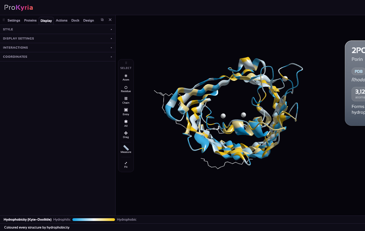
Even before adding a membrane, you can see a band of hydrophobic residues running around the outside of the barrel. That is where the lipids will be.
2. Find the aromatic girdle
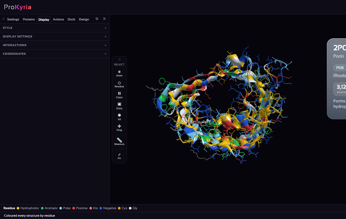
Now colour by Residue type, still with nothing selected. Aromatic residues (phenylalanine, tryptophan and tyrosine) turn green, and porin's sixteen tyrosines and its tryptophan form two rings, one at each edge of the yellow band. This aromatic girdle is typical of membrane proteins: the flat rings sit where the oily core of the membrane meets the lipids' heads and the water, and anchor the protein at the right depth.
To see them better, select the whole entry (Entry on the tool wand, then click it), turn on Stick as well as Ribbon, and colour by Residue type again.
3. Add the bilayer
Open Actions and press Add Lipid Bilayer. ProKyria searches for the orientation and depth that best bury the protein's hydrophobic, outward-facing residues, with a bonus for aromatic residues at the edges, and builds a bilayer there, with lipids cut away where the protein is.
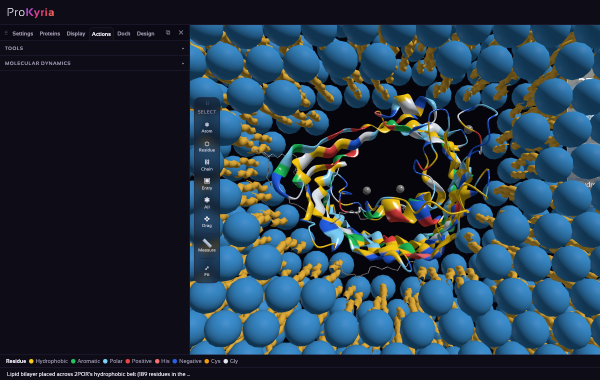
The status bar, and the step in the Analysis log, report the result: here it found the belt, with 189 residues in the membrane core. If a protein has no convincing belt, ProKyria says it looks soluble and places the bilayer at its most hydrophobic face instead.
4. Check the fit from the side
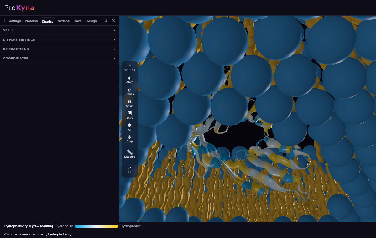
Colour by Hydrophobicity again and rotate the view to look along the membrane. The yellow band should fill the lipid tails exactly, with the green aromatic rings at the level of the heads. Check the edges: hydrophilic sky-blue residues inside the core, or yellow ones far outside it, would mean a poor placement.
5. Taking it further
- The bilayer is an entry of its own, Lipid bilayer in Loaded Entries: select it to colour or style it, for example by Element or Solid, or remove it like any entry. It's 240 Å across, made of POPC with a quarter cholesterol. If you move the protein, press Add Lipid Bilayer again to rebuild it in the new place.
- Turn on Display › Interactions, with Hydrogen bonds and only within a chain, to see the bonds between neighbouring strands that hold the barrel together.
- Try a helical membrane protein, such as bacteriorhodopsin (
1C3W), to compare its belt with porin's.
More detail: how membrane placement works.