Not every protein has an experimental structure. But with modern structure prediction, a sequence is often enough for a good model. ProKyria uses ESMFold, a protein language model from Meta AI, to predict a structure from one sequence, typically in seconds. We'll fold ubiquitin, read how sure the model is, and mark the residues that do ubiquitin's job.
1. Enter the sequence
Open the Predict section of the Proteins tab. Paste a sequence into the box: one-letter codes, plain or in FASTA format. You can also use Load sequence from file, drop a FASTA file onto the box, click one of the Examples, or search for a protein by name with Search at the top. We'll use ubiquitin, 76 residues.
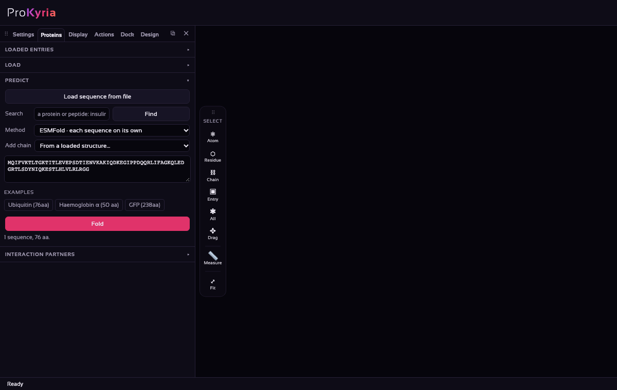
Leave the method on ESMFold · each sequence on its own. If you paste several sequences, each one is folded separately. ESMFold co-fold instead folds them together as one complex, which is useful for heterodimers. Use Add chain to take a chain from a structure you've already loaded.
2. Fold it and follow the chain
Press Fold. The sequence is sent to the ESM Metagenomic Atlas service, and the model comes back and loads like any other structure, marked Pred. in the side panel.
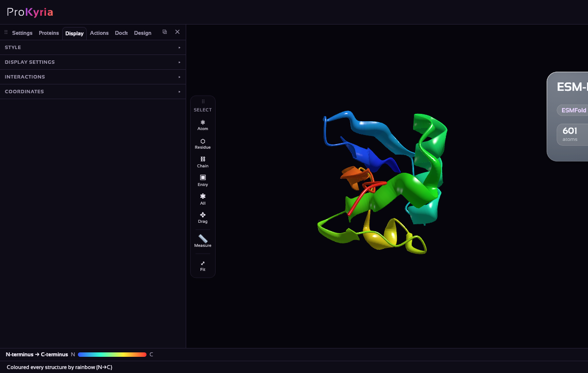
In Display › Style, choose Ribbon and colour by Rainbow: the chain runs from blue at the N-terminus to red at the C-terminus. Follow it: a β-hairpin, a helix packed against the sheet, then more strands, the compact β-grasp fold, with the red tail sticking out at the end.
3. Read the confidence
Now colour by pLDDT, the model's own estimate of how reliable each residue is, from 0 to 100.
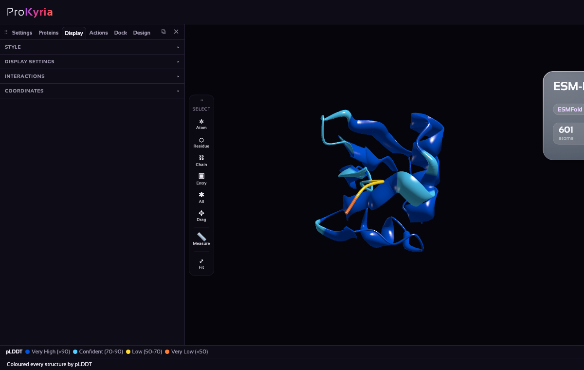
- Very high (> 90): dark blue. Usually accurate down to side-chain level.
- Confident (70–90): light blue. The backbone is generally right.
- Low (50–70): yellow. Treat with caution.
- Very low (< 50): orange. Often a disordered region, not a real structure.
Ubiquitin is a compact, well-studied fold, and almost all of the model is very high confidence. Only the flexible tail at the end is less certain, which is just what you'd expect.
4. Mark the working parts
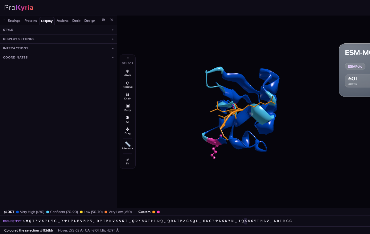
Ubiquitin tags other proteins: its C-terminal Gly76 is joined to a lysine on the target, and further ubiquitins are chained on through its own lysines, Lys48 (a signal for destruction) and Lys63 (a signal for signalling and repair). Turn on the sequence bar (Display › Display settings › Amino acid sequence):
- Click L73 and Shift-click G76 to select the tail. Show it as Stick with the Solid colour chip set to orange.
- Click K48 and Ctrl/⌘-click K63. Show them as Stick too, coloured Solid magenta.
The two lysines point out from different faces of the fold, so chains linked through them have different shapes, and the cell reads them as different signals. The tail is where the model is least sure, because in reality it is flexible.
Tips and limits
- ESMFold works on single sequences, so it needs no alignment. For some proteins, alignment-based methods such as AlphaFold2 do better. For proteins already in UniProt, load the precomputed AlphaFold model from Load a Structure.
- The service accepts sequences up to about 400 residues.
- Your sequence is sent to an outside service. Don't fold confidential sequences.
More detail: structure prediction with ESMFold.